Sketch And Label Of A Cross Section Of A Long Bone ~ Anatomy Wikipedia. This is the long central shaft. This is an online quiz called long bone anatomy. A = epiphysis b = diaphysis c = articular cartilage d = periosteum f = compact bone g = medullary cavity (yellow marrow) h = endosteum j = epiphyseal line (growth plate) coloring worksheet for this image. This problem has been solved! There is a printable worksheet available for download here so you can take the quiz with pen and paper.
Sketch and label a cross. Related posts of cross section of a long bone bone test anatomy and physiology. Human back muscles and bones 12 photos of the human back muscles and bones human back muscles and bones, bone, human back muscles and bones. Label lines should not cross ; A flat bone is characterized by parallel surfaces of figure figure observe a slide preparation labeled ground bone;
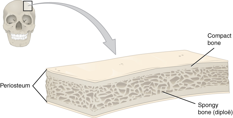
Flat Bone Wikipedia from upload.wikimedia.org A long bone has a shaft and 2 ends. The diaphysis and the epiphysis.the diaphysis is the tubular shaft that runs between the proximal and distal ends of the bone. This problem has been solved! External circumferential lamellae, osteon, central canal, perforating canals, lacuna, canaliculi, concentric lamellae. There is a printable worksheet available for download here so you can take the quiz with pen and paper. Marks should be deducted for shading or colouring. The periosteum contains many strong collagen fibers that are used to firmly anchor tendons and muscles to the bone for movement. Click on the tags below to find other quizzes on the same subject.
The hollow region in the diaphysis is called the medullary cavity, which is filled.
The diaphysis and the epiphysis. Exploring the microscopic anatomy of bone 1. Sketch and label a cross. The hollow region in the diaphysis is called the medullary cavity, which is filled. The original can be viewed here: Human back muscles and bones 12 photos of the human back muscles and bones human back muscles and bones, bone, human back muscles and bones. Then, fill in the table below to describe each. As shown in figure 2. Long bones have a thick outside layer of compact bone and an inner medullary cavity containing bone marrow. Related posts of cross section of a long bone bone test anatomy and physiology. The diaphysis is the tubular shaft that runs between the proximal and distal ends of the bone. Forms the larger rounded ends of long bones. Get premium, high resolution news photos at getty images
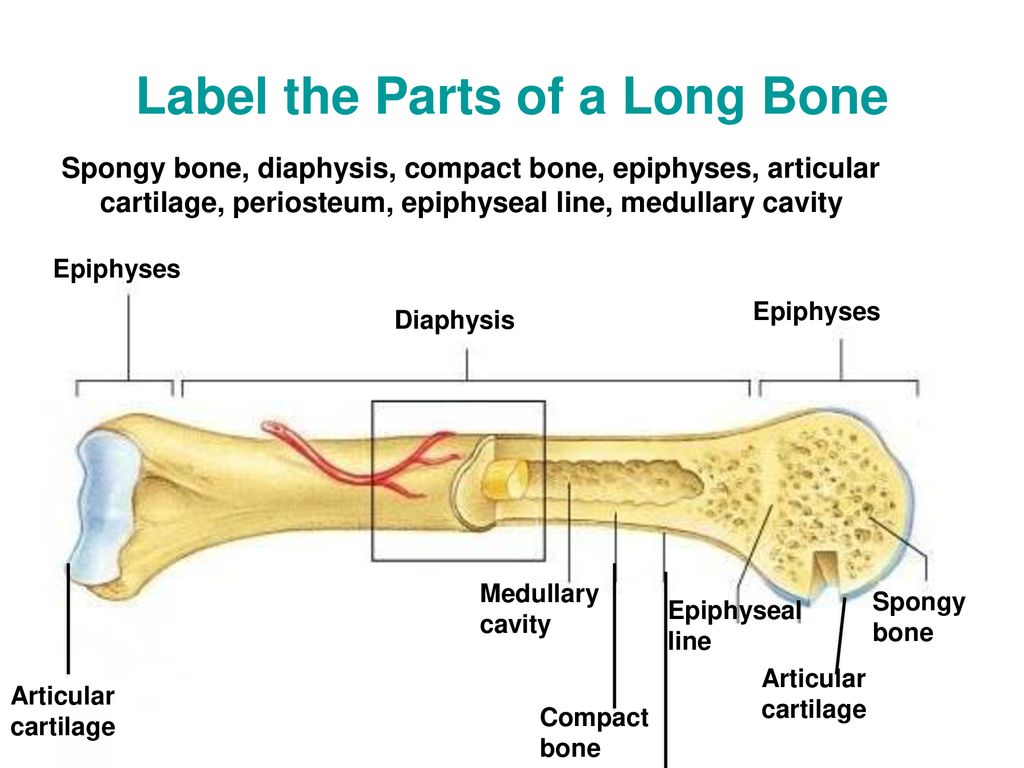
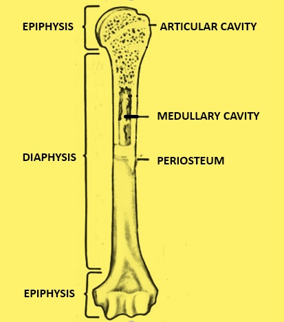
The diaphysis and the epiphysis. Draw and label a longitudinal section of a long bone. The structure of a long bone allows for the best visualization of all of the parts of a bone (figure 6.7). Bone cross section + long bone. The structure of a long bone allows for the best visualization of all of the parts of a bone ().a long bone has two parts:
Skeletal System Ppt Download from slideplayer.com The outside of a bone is covered in a thin layer of dense irregular connective tissue called the periosteum. (do not copy and paste a picture from the text or internet.) Area between the diaphysis and epiphysis at both ends of the bone. Forms the larger rounded ends of long bones. The structure of a long bone consists of several sections:. The structure of a long bone allows for the best visualization of all of the parts of a bone ().a long bone has two parts: Label the parts of a long bone. Draw and label a longitudinal section of a long bone.
Anatomy of shoulder 12 photos of the anatomy of shoulder anatomy of nerves in shoulder, anatomy of posterior shoulder dislocation, anatomy of right shoulder, anatomy of shoulder labrum tear, anatomy of the shoulder games, human anatomy, anatomy of nerves in shoulder, anatomy of posterior shoulder dislocation, anatomy of.
A long bone has a shaft and 2 ends. Looking at a bone in cross section, there are several distinct layered regions that make up a bone. The walls of the diaphysis are composed of dense and hard compact bone. The diaphysis and the epiphysis. There is a printable worksheet available for download here so you can take the quiz with pen and paper. (do not copy and paste a picture from the text or internet.) 3. The periosteum contains many strong collagen fibers that are used to firmly anchor tendons and muscles to the bone for movement. Click on the tags below to find other quizzes on the same subject. The ends of a long bone contain spongy bone and an epiphyseal line. Label the haversian canal, osteocyte (mature bone cell) in lacuna, and canaliculi. Related posts of compact bone diagram labeled anatomy of shoulder. Smartdraw includes 1000s of professional healthcare and anatomy chart templates that you can modify and make your own. The structure of a long bone allows for the best visualization of all of the parts of a bone ().a long bone has two parts:
The compact bone is made up of osteon. Exploring the microscopic anatomy of bone 1. Bones at college of technology at farmingdale (farmingdale state. A long bone has two parts: This problem has been solved!
Sketch And Label Of A Cross Section Of A Long Bone Long Bone Cross Section Worksheet Teaching Resources As The Names Suggest Compact Bone Looks Compact And The Spongy Bone from d2nchlq0f2u6vy.cloudfront.net Two types of bone tissues in cross section of a long bone : The structure of a long bone consists of several sections:. A = epiphysis b = diaphysis c = articular cartilage d = periosteum f = compact bone g = medullary cavity (yellow marrow) h = endosteum j = epiphyseal line (growth plate) coloring worksheet for this image. A long bone has a shaft and 2 ends. Compact bone is the outer layer and the spongy bone forms the inner layer. The diaphysis and the epiphysis. Then, fill in the table below to describe each. (do not copy and paste a picture from the text or internet.) 3.
A long bone has two parts:
The diaphysis and the epiphysis. Sketch a longitudinal section through a long bone and label the following structures de epiphysim ercavi periosteum, co p pseen, compact bune.no red bone marrow, and yellow bone marrow he provides a epiphysis riedullary activity 4: Marks should be deducted for shading or colouring. Two types of bone tissues in cross section of a long bone : Label the parts of a long bone. Learners should accurately draw a long bone, resembling that in figure 6.24. A flat bone is characterized by parallel surfaces of figure figure observe a slide preparation labeled ground bone; Click on the tags below to find other quizzes on the same subject. The ends of a long bone contain spongy bone and an epiphyseal line. In these labeled examples, a human femur is represented without identifying many of the unique characteristics that help differentiate the femur bone from other bones in the human body. Then, fill in the table below to describe each. Bone matrix and cells bone matrix osseous tissue is a connective tissue and like all connective tissues contains relatively few cells and large amounts of extracellular matrix. The structure of a long bone allows for the best visualization of all of the parts of a bone ().a long bone has two parts: